Using ultrasound imaging in filler injection procedures brings a new level of precision to aesthetic medicine. This advancement enhances both safety and accuracy in achieving the desired aesthetic outcomes. In this article, we will explore ultrasound imaging, its role in filler injections, and the benefits it offers to practitioners and patients in ensuring a safer and more precise procedure.
Ultrasound Imaging in Aesthetic Medicine
Ultrasound imaging, a technique widely recognised in the medical field for its diagnostic capabilities, has found a significant place in aesthetic medicine over recent years. The technology, known for its non-invasive nature and real-time imaging capabilities, has become a cornerstone in enhancing the safety and accuracy of aesthetic procedures.
The essence of ultrasound imaging transcends the traditional touch or palpation techniques, which were once staples in the administration of dermal fillers. It introduces a paradigm where practitioners can now visualise the underlying anatomical structures, including vascular anatomy, in real-time. This transition has not only reduced the risks associated with blind injections but also augments the precision and safety with which dermal fillers are administered.
What is Ultrasound Imaging?
Ultrasound imaging, or ultrasonography, is a diagnostic imaging technique that uses high-frequency sound waves to create images of the internal structures of the body. Unlike X-rays, it doesn’t involve radiation, making it a safer alternative for imaging. When applied to the field of aesthetic medicine, particularly in filler injections, ultrasound imaging becomes a powerful tool that aids in real-time visualisation of the treatment area.
This enables practitioners to visualise the needle’s path and filler placement, fostering a more accurate and safer procedure. Additionally, ultrasound imaging provides an opportunity for practitioners to educate patients by showing them the images and explaining the procedure, thus enhancing the overall patient experience.
How Does Ultrasound Contribute to More Consistent and Safer Filler Results?
This technology brings greater accuracy and safety to the administration of dermal fillers, which are used to restore volume, smooth lines, and enhance facial contours. Here are some of the noteworthy advantages of employing ultrasound guidance during dermal filler injections:
- Precision: Ultrasound imaging provides real-time, high-definition images of the underlying anatomical structures, enabling practitioners to precisely target the desired injection sites. This precision is paramount, especially when working in proximity to critical structures such as blood vessels and nerves.
- Safety: By visualising the anatomy beneath the skin, practitioners can avoid inadvertently injecting fillers into blood vessels, a complication that can lead to serious adverse events. The real-time imaging allows for immediate corrective actions if any misplacement is observed, significantly enhancing the safety profile of dermal filler procedures.
- Efficiency and Effectiveness: The use of ultrasound can potentially reduce the amount of filler material needed to achieve the desired result, thanks to the precise placement. Additionally, it allows for immediate verification of results, saving time and ensuring the effectiveness of the treatment.
- Education and Skill Enhancement: Ultrasound guidance serves as an invaluable educational tool for practitioners. It provides immediate feedback on the accuracy of filler placement, facilitating a better understanding and improvement of injection techniques over time.
- Patient Confidence: Knowing that an advanced imaging technology is being used can enhance patient confidence in the procedure and the practitioner. It sets a higher standard of care, aligning with the expectations of an informed patient seeking safe and effective aesthetic treatments.
- Reduced Complication Management: In the rare event of complications, ultrasound imaging can be used to accurately locate and quantify any residual filler material, aiding in the formulation of a management or corrective plan.
The incorporation of ultrasound technology in dermal filler injections not only elevates the standard of care but also significantly contributes to achieving better aesthetic outcomes with a higher safety margin. It addresses the crucial need for accurate filler placement and the avoidance of critical structures like blood vessels, which is pivotal in reducing complications.
The Procedure of Ultrasound Imaging Filler Injections
Preparation
The initial step toward a successful ultrasound imaging filler injection procedure is preparation. During this phase, the treatment area is thoroughly cleaned and prepped to ensure it is free from any impurities that may hinder the procedure or cause infections. Additionally, to enhance the comfort of the patient, a topical anaesthetic may be applied to numb the area, making the procedure less discomforting.
Ultrasound Imaging
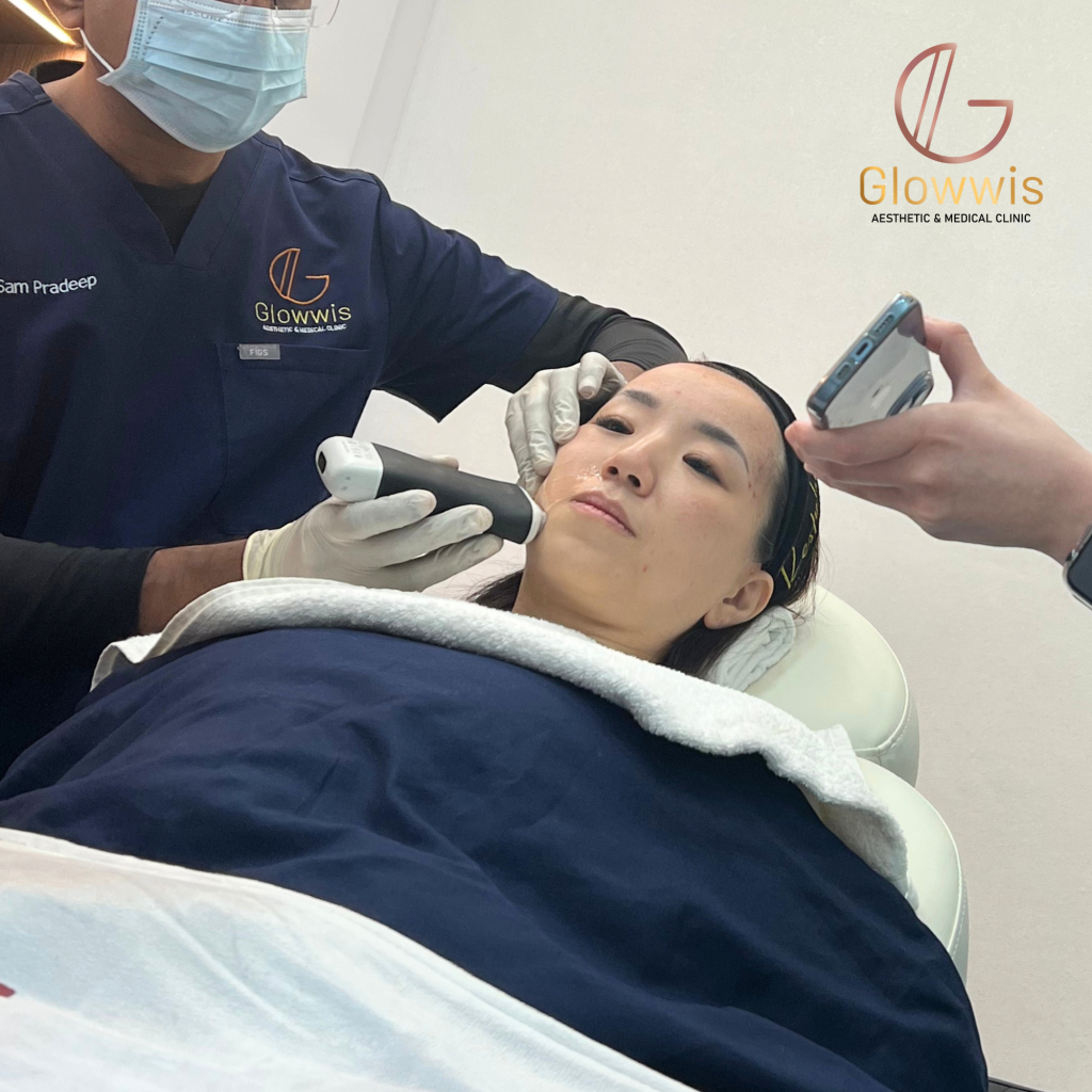
Following preparation, the practitioner transitions to the ultrasound imaging phase. Utilising a handheld ultrasound device, they scan the treatment area meticulously. This crucial step aids in identifying the location and depth of blood vessels and other critical structures, thereby providing a clear anatomical roadmap for the practitioner. This information is vital as it significantly contributes to the safety and accuracy of the filler injection process.
Marking the Injection Sites
Based on the insights garnered from the ultrasound imaging, the practitioner marks the precise injection sites on the skin. This marking acts as a guideline to ensure accurate filler placement, significantly reducing the risks associated with blind injections.
Administering the Filler
With a clear image , the practitioner moves to administering the filler phase. Using a syringe, the practitioner administers the filler, continually monitoring the ultrasound imaging. This continuous monitoring ensures accurate filler placement and aids in avoiding critical structures that, if disturbed, could lead to complications.
Real-Time Monitoring
Ultrasound imaging provides real-time feedback, enabling the practitioner to make necessary adjustments to the needle position, ensuring accurate filler placement, and confirming that the filler is deposited in the desired location.
Post-Procedure Assessment
Post-procedure, an assessment of the results is conducted. The practitioner scrutinises the treated area, and if necessary, further adjustments can be made to achieve the desired aesthetic outcome. Moreover, the ultrasound may be utilised again to confirm the results and ensure that no complications have occurred, affirming the success of the procedure.
Aftercare
Lastly, the procedure transitions to the aftercare phase. Patients are provided with detailed aftercare instructions to ensure the best possible healing and results. This guidance is crucial as proper aftercare significantly contributes to the success and satisfaction with the ultrasound imaging filler injections procedure.
Each step of this procedure underscores the indispensable role of ultrasound imaging in enhancing the safety, accuracy, and overall success of filler injections.
The Future of Ultrasound Imaging Filler Injections
The integration of ultrasound guidance in dermal filler injections is increasingly being adopted, potentially setting a new benchmark in the field. As the technology evolves, improvements in imaging resolution and user-friendly devices are expected. Additionally, specialised training programmes may be introduced to ensure practitioners develop the necessary expertise in this technique.
Glowwis Aesthetic & Medical Clinic already utilises ultrasound guidance for filler injections. As more patients become informed about the benefits of ultrasound guidance, the demand for such progressive practices is likely to increase. Ongoing research and collaboration between medical practitioners and technology providers may further facilitate the use of ultrasound-guided filler injections, contributing to its role in modern aesthetic treatments.
Conclusion
The use of ultrasound imaging in dermal filler procedures represents a notable development that enhances safety, precision, and aesthetic outcomes. As technology continues to improve, incorporating such advancements is key to refining practice standards in aesthetic medicine, ultimately benefiting both practitioners and patients.